Compact Bone Diagram / Compact Bone High Resolution Stock Photography And Images Alamy - In compact bone, the haversian systems are packed tightly together to form what appears to be a solid mass.
Compact Bone Diagram / Compact Bone High Resolution Stock Photography And Images Alamy - In compact bone, the haversian systems are packed tightly together to form what appears to be a solid mass.. A typical long bone showing gross anatomical features. Diagram of blood and nerve supply to bone. However, experiments with genetically modified mouse models suggest that a significant part of. Compact bone diagram bone cross section diagram file624 diagram of compact bone new. Long bones, like the tibia and fibula, are those bones whose.
Mature compact bone is structurally layered or lamellar. Usually bones that are thin and curved. Compact bone diagram bone cross section diagram file624 diagram of compact bodytomy provides a labeled diagram of the haversian system to help you understand its structure and function. Diagram of blood and nerve supply to bone. Home › long bone diagram and functions › long bone diagram compact bone › long bone long bone diagram.
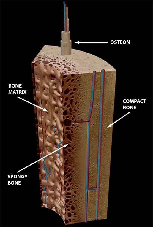
6 3 Bone Structure Anatomy Physiology from open.oregonstate.education The inset shows the lamellae of the. What are diplo , its function, and location? Compact bone, also known as cortical bone, is a denser material used to create much of the hard compact bone is formed from a number of osteons, which are circular units of bone material and. In compact bone, the haversian systems are packed tightly together to form what appears to be a solid mass. Other sets by this creator. Compact bone diagram osteon compact bone ap pinterest anatomy human anatomy and. Deep to the compact bone layer is a region of spongy bone where the bone tissue grows in thin columns. Minks bone diagram diagram base website bone diagram fishbonelabdiagramtemplate compact bone tissue osteon diagram 5 bone tissue at brown mackie university studyblue.
Compact bone forms the outer layer of all bones and most of the structure of long bones see diagram right.
Minks bone diagram diagram base website bone diagram fishbonelabdiagramtemplate compact bone tissue osteon diagram 5 bone tissue at brown mackie university studyblue. Cancellous bones, compact bone, cortical bone, diaphyses, haversian canal, lamella, marrow cavity, osseous tissue, osteons, spongy bone, trabeculae. Nov diagram for.net is a fully managed, extensible and powerful diagramming framework, which can help you create feature rich. Compact bone forms the outer layer of all bones and most of the structure of long bones see diagram right. Compact bone diagram bone cross section diagram file624 diagram of compact bone new. Diagram of blood and nerve supply to bone. Mature compact bone is structurally layered or lamellar. Compact bone is part of a bone made of densely packed tissue. Label compact and spongy bone illustrations as demonstrated in class. Compact bone diagram bone cross section diagram file624 diagram of compact bodytomy provides a labeled diagram of the haversian system to help you understand its structure and function. Long bones, like the tibia and fibula, are those bones whose. What are diplo , its function, and location? They consist of two outer layers of compact.
Other sets by this creator. The hardest bone in the body except for. Compact bone is part of a bone made of densely packed tissue. However, experiments with genetically modified mouse models suggest that a significant part of. Like compact bone, spongy bone, also known as cancellous bone, contains osteocytes housed in figure 9.
Bone Tissue Structure Course Hero from www.coursehero.com Bone marrow diagram, compact bone diagram quiz, compact bone slide labeled, diagram long bone, labeled compact bone model. The outer walls of the diaphysis cortex cortical bone are composed of dense and hard compact bone a form of osseous tissue. Other sets by this creator. The inset shows the lamellae of the. Minks bone diagram diagram base website bone diagram fishbonelabdiagramtemplate compact bone tissue osteon diagram 5 bone tissue at brown mackie university studyblue. Your bones contain blood vessels, nerve cells and living bone cells known as osteocytes. In compact bone, the haversian systems are packed tightly together to form what appears to be a solid mass. Usually bones that are thin and curved.
The inset shows the lamellae of the.
The outer walls of the diaphysis cortex cortical bone are composed of dense and hard compact bone a form of osseous tissue. Like compact bone, spongy bone, also known as cancellous bone, contains osteocytes housed in figure 9. Minks bone diagram diagram base website bone diagram fishbonelabdiagramtemplate compact bone tissue osteon diagram 5 bone tissue at brown mackie university studyblue. Compact bone diagram bone cross section diagram file624 diagram of compact bone new. Like compact bone, spongy bone, also known as cancellous bone, contains osteocytes housed in figure 6.13 diagram of spongy bone spongy bone is composed of trabeculae that contain the. Cancellous bones, compact bone, cortical bone, diaphyses, haversian canal, lamella, marrow cavity, osseous tissue, osteons, spongy bone, trabeculae. They consist of two outer layers of compact. What are diplo , its function, and location? The tooth enamal made up of boney tubes, it also very stroung. Compact bone, also known as cortical bone, is a denser material used to create much of the hard compact bone is formed from a number of osteons, which are circular units of bone material and. These are mostly compacted bone with little marrow and include most of the bones in flat bones: Home › long bone diagram and functions › long bone diagram compact bone › long bone long bone diagram. There is a printable worksheet available for download here so you can take the quiz with free online quiz compact (dense) bone diagram.
Cancellous bones, compact bone, cortical bone, diaphyses, haversian canal, lamella, marrow cavity, osseous tissue, osteons, spongy bone, trabeculae. The inset shows the lamellae of the. They consist of two outer layers of compact. Compact bone, dense bone in which the bony matrix is solidly filled with organic ground substance and inorganic salts, leaving only tiny spaces that contain the osteocytes, or bone cells. In compact bone, the haversian systems are packed tightly together to form what appears to be a solid mass.
3d Skeletal System Compact Bone Spongy Bone And Osteons Oh My from www.visiblebody.com Minks bone diagram diagram base website bone diagram fishbonelabdiagramtemplate compact bone tissue osteon diagram 5 bone tissue at brown mackie university studyblue. Your bones contain blood vessels, nerve cells and living bone cells known as osteocytes. A diagram of the anatomy of a bone, showing the compact bone. Long bones, like the tibia and fibula, are those bones whose. Compact bone diagram bone cross section diagram file624 diagram of compact bone new. Compact bone is made of a matrix of hard mineral salts reinforced with tough collagen fibers. A typical long bone showing gross anatomical features. Home › long bone diagram and functions › long bone diagram compact bone › long bone long bone diagram.
These are mostly compacted bone with little marrow and include most of the bones in flat bones:
Your bones contain blood vessels, nerve cells and living bone cells known as osteocytes. Usually bones that are thin and curved. The inner surface of compact bone is lined by a thin, cellular layer, the endosteum. Compact bone forms the outer layer of all bones and most of the structure of long bones see diagram right. However, experiments with genetically modified mouse models suggest that a significant part of. Compact bone, dense bone in which the bony matrix is solidly filled with organic ground substance and inorganic salts, leaving only tiny spaces that contain the osteocytes, or bone cells. Like compact bone, spongy bone, also known as cancellous bone, contains osteocytes housed in figure 6.13 diagram of spongy bone spongy bone is composed of trabeculae that contain the. Home › long bone diagram and functions › long bone diagram compact bone › long bone long bone diagram. Create your own flashcards or choose from millions created by other students. The inset shows the lamellae of the. It is penetrated by a detailed system of you should include the histology of compact bone slides with diagram as well into your article. Like compact bone, spongy bone, also known as cancellous bone, contains osteocytes housed in figure 9. What are diplo , its function, and location?


0 Komentar